File:MRI pilocytic astrocytoma 5.jpg
Jump to navigation
Jump to search
Size of this preview: 256 × 599 pixels. Other resolution: 260 × 608 pixels.
Original file (260 × 608 pixels, file size: 46 KB, MIME type: image/jpeg)
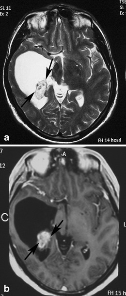
Classical appearance of a hemispheric pilocytic astrocytoma in a 45-year-old man. a An axial T2-weighted image demonstrates a hyperintense cystic component and a less hyperintense solid nodule within the lateral ventricle (arrows). b A contrast-enhanced axial T1-weighted image reveals intense enhancement of the solid nodule (arrows) and lack of enhancement of the cystic portion.
File history
Click on a date/time to view the file as it appeared at that time.
| Date/Time | Thumbnail | Dimensions | User | Comment | |
|---|---|---|---|---|---|
| current | 20:53, 29 October 2015 | | 260 × 608 (46 KB) | Sujit Routray (talk | contribs) | Classical appearance of a hemispheric pilocytic astrocytoma in a 45-year-old man. '''a''' An axial T2-weighted image demonstrates a hyperintense cystic component and a less hyperintense solid nodule within the lateral ventricle (arrows). '''b''' A cont... |
| 20:53, 29 October 2015 |  | 260 × 608 (46 KB) | Sujit Routray (talk | contribs) | Classical appearance of a hemispheric pilocytic astrocytoma in a 45-year-old man. '''a''' An axial T2-weighted image demonstrates a hyperintense cystic component and a less hyperintense solid nodule within the lateral ventricle (arrows). '''b''' A cont... |
You cannot overwrite this file.
File usage
There are no pages that use this file.