File:MRI pilocytic astrocytoma 4.jpg
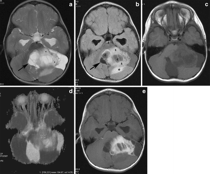
Pilocytic astrocytoma of the left cerebellar hemisphere with a solid and cystic component severely compressing the fourth ventricle and the medulla, producing obstructive hydrocephalus. a An axial T2-weighted image shows a cystic, hyperintense mass with a less intense solid component (asterisks) compressing the fourth ventricle (arrow). b On a FLAIR image, the cystic component (c) shows low signal intensity, which is higher than that of the CSF and the solid component (s) shows high signal intensity. Note the low signal of the compressed fourth ventricle (arrow) and the peritumoral edema (e). c On this T1-weighted image, the solid components appear hypointense compared with grey matter. d The solid component shows high values on an ADC map. e Intense enhancement of solid components is depicted after the administration of a paramagnetic agent.
File history
Click on a date/time to view the file as it appeared at that time.
| Date/Time | Thumbnail | Dimensions | User | Comment | |
|---|---|---|---|---|---|
| current | 17:49, 29 October 2015 | | 709 × 589 (85 KB) | Sujit Routray (talk | contribs) | Pilocytic astrocytoma of the left cerebellar hemisphere with a solid and cystic component severely compressing the fourth ventricle and the medulla, producing obstructive hydrocephalus. '''a''' An axial T2-weighted image shows a cystic, hyperintense ma... |
| 17:48, 29 October 2015 |  | 709 × 589 (85 KB) | Sujit Routray (talk | contribs) | Pilocytic astrocytoma of the left cerebellar hemisphere with a solid and cystic component severely compressing the fourth ventricle and the medulla, producing obstructive hydrocephalus. '''a''' An axial T2-weighted image shows a cystic, hyperintense ma... |
You cannot overwrite this file.
File usage
There are no pages that use this file.