File:HIV pathophysiology01.jpeg
Original file (700 × 840 pixels, file size: 77 KB, MIME type: image/jpeg)
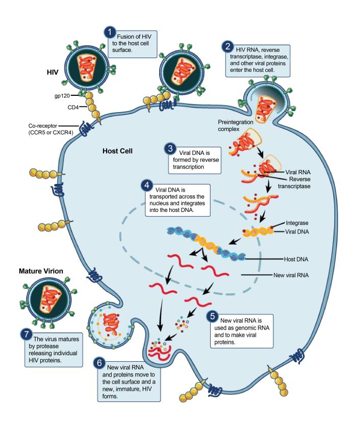
Produced by the National Institute of Allergy and Infectious Diseases (NIAID), this illustration depicts the human immunodeficiency virus (HIV) replication cycle involving a host cell within the human body, beginning with a HIV virion attaching to the host cell wall, dumping its contents into the matrix of the host cell, conversion of the viral RNA to viral DNA, which combines with the host cell DNA, leading to the creation of new viral RNA, which migrates to the host cell periphery, and forms a vacuole around the new viral RNA using the host cell wall, thereafter, budding off as a newly-created mature HIV virion. Please see the Flickr link below for additional NIAID photomicrographs of various microbes.
File history
Click on a date/time to view the file as it appeared at that time.
| Date/Time | Thumbnail | Dimensions | User | Comment | |
|---|---|---|---|---|---|
| current | 16:30, 20 November 2014 | | 700 × 840 (77 KB) | Jesus Hernandez (talk | contribs) | Produced by the National Institute of Allergy and Infectious Diseases (NIAID), this illustration depicts the human immunodeficiency virus (HIV) replication cycle involving a host cell within the human body, beginning with a HIV virion attaching to the ... |
You cannot overwrite this file.
File usage
The following page uses this file: