File:CT scan of pilocytic astrocytoma.jpg
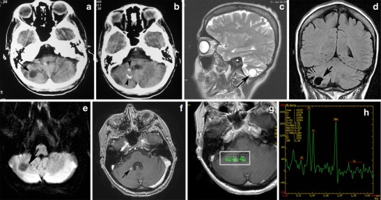
A pilocytic astrocytoma of the right cerebellar hemisphere incidentally discovered during staging in a 45-year-old woman with breast cancer. a An axial CT image shows a well-marginated cyst in the right cerebellar hemisphere (arrow). b An axial CT image in a higher level shows fleck-like calcifications (arrowheads). On sagittal T2-weighted ('c), coronal FLAIR (d), and axial diffusion-weighted (e) images, the cyst follows cerebrospinal fluid intensity (arrows). This axial post-contrast T1-weighted image (f) depicts an enhancing focus (arrow). On MRS centred on an enhancing nodule with TE 144-ms (g, h), there is mild elevation of choline/creatine, mild reduction of NAA/vreatine, and a small lactate peak.
File history
Click on a date/time to view the file as it appeared at that time.
| Date/Time | Thumbnail | Dimensions | User | Comment | |
|---|---|---|---|---|---|
| current | 14:44, 29 October 2015 | | 547 × 288 (59 KB) | Sujit Routray (talk | contribs) | A pilocytic astrocytoma of the right cerebellar hemisphere incidentally discovered during staging in a 45-year-old woman with breast cancer. '''a''' An axial CT image shows a well-marginated cyst in the right cerebellar hemisphere (arrow). '''b''' An a... |
| 14:43, 29 October 2015 |  | 547 × 288 (59 KB) | Sujit Routray (talk | contribs) | A pilocytic astrocytoma of the right cerebellar hemisphere incidentally discovered during staging in a 45-year-old woman with breast cancer. '''a''' An axial CT image shows a well-marginated cyst in the right cerebellar hemisphere (arrow). '''b''' An a... |
You cannot overwrite this file.
File usage
There are no pages that use this file.