File:CT Juvenile Pilocytic Astrocytoma.jpg
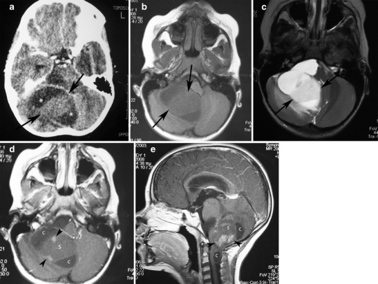
Juvenile pilocytic astrocytoma of the cerebellum in a 5-year-old girl. a Post-contrast CT shows a cystic lesion involving the right cerebellar hemisphere with a hypodense cystic component (asterisks) and a large, solid, isodense component (arrows). b On axial T1-weighted image, the solid component is homogeneous and hypointense compared with the grey matter (arrows). c On T2-weighted images, the solid component appears hyperintense compared with the grey matter and slightly hypointense compared with the cerebrospinal fluid (arrows). Post-contrast axial (d) and sagittal (e) images demonstrate heterogeneous enhancement of the solid component of the mass with areas that remain unenhanced (s) and areas with nodular enhancement (arrowheads). Note the cystic components (c)
File history
Click on a date/time to view the file as it appeared at that time.
| Date/Time | Thumbnail | Dimensions | User | Comment | |
|---|---|---|---|---|---|
| current | 14:37, 29 October 2015 | | 547 × 411 (64 KB) | Sujit Routray (talk | contribs) | Juvenile pilocytic astrocytoma of the cerebellum in a 5-year-old girl. '''a''' Post-contrast CT shows a cystic lesion involving the right cerebellar hemisphere with a hypodense cystic component (asterisks) and a large, solid, isodense component (arrows... |
| 14:36, 29 October 2015 |  | 547 × 411 (64 KB) | Sujit Routray (talk | contribs) | Juvenile pilocytic astrocytoma of the cerebellum in a 5-year-old girl. '''a''' Post-contrast CT shows a cystic lesion involving the right cerebellar hemisphere with a hypodense cystic component (asterisks) and a large, solid, isodense component (arrows... |
You cannot overwrite this file.
File usage
There are no pages that use this file.