File:Amebiasis07.jpeg
Jump to navigation
Jump to search
No higher resolution available.
Amebiasis07.jpeg (700 × 525 pixels, file size: 42 KB, MIME type: image/jpeg)
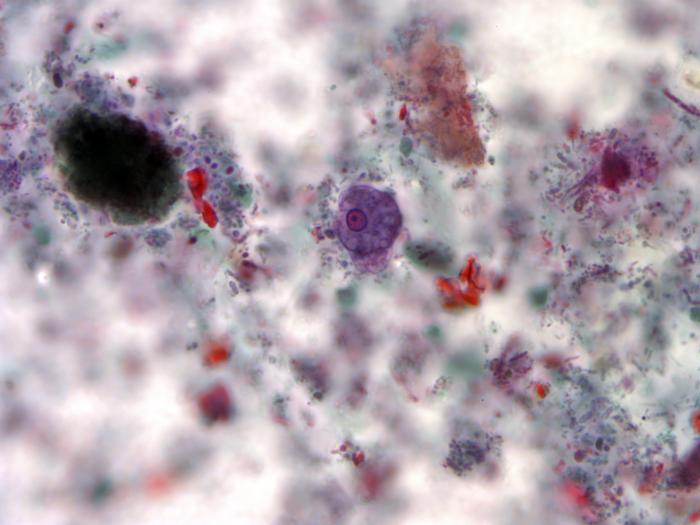
Using a trichrome stain, this photomicrograph depicted a trophozoite of the single-celled parasite, Entamoeba histolytica. Stained purple, the trophozoite, see here in the center of the micrograph, is one of the life cycle phases through which a protozoan organism passes as it matures, and is the active-feeding phase of its growth. The other particulates surrounding the trophozoite represent debris from the slide specimen.
File history
Click on a date/time to view the file as it appeared at that time.
| Date/Time | Thumbnail | Dimensions | User | Comment | |
|---|---|---|---|---|---|
| current | 16:47, 20 November 2014 | | 700 × 525 (42 KB) | Jesus Hernandez (talk | contribs) | Using a trichrome stain, this photomicrograph depicted a trophozoite of the single-celled parasite, Entamoeba histolytica. Stained purple, the trophozoite, see here in the center of the micrograph, is one of the life cycle phases through which a protoz... |
You cannot overwrite this file.
File usage
The following file is a duplicate of this file (more details):
The following page uses this file: