Extensor hallucis longus muscle
Overview
The Extensor hallucis longus is a thin muscle, situated between the Tibialis anterior and the Extensor digitorum longus that functions to extend the big toe, dorsiflex the foot, and assists with foot inversion.
It arises from the anterior surface of the fibula for about the middle two-fourths of its extent, medial to the origin of the Extensor digitorum longus; it also arises from the interosseous membrane to a similar extent.
The anterior tibial vessels and deep peroneal nerve lie between it and the Tibialis anterior.
The fibers pass downward, and end in a tendon, which occupies the anterior border of the muscle, passes through a distinct compartment in the cruciate crural ligament, crosses from the lateral to the medial side of the anterior tibial vessels near the bend of the ankle, and is inserted into the base of the distal phalanx of the great toe.
Opposite the metatarsophalangeal articulation, the tendon gives off a thin prolongation on either side, to cover the surface of the joint.
An expansion from the medial side of the tendon is usually inserted into the base of the proximal phalanx.
Variations
Occasionally united at its origin with the Extensor digitorum longus.
Extensor ossis metatarsi hallucis, a small muscle, sometimes found as a slip from the Extensor hallucis longus, or from the Tibialis anterior, or from the Extensor digitorum longus, or as a distinct muscle; it traverses the same compartment of the transverse ligament with the Extensor hallucis longus.
Additional images
-
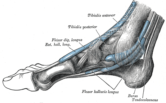 The mucous sheaths of the tendons around the ankle. Medial aspect.
 -
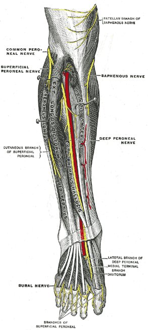 Deep nerves of the front of the leg.
 -
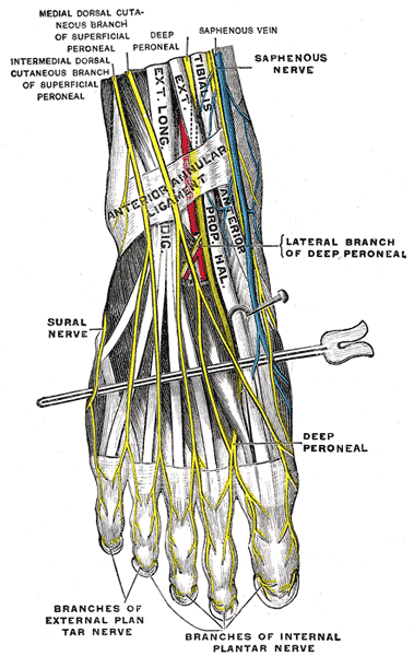 Nerves of the dorsum of the foot.


External links
- Template:GPnotebook
- Template:SUNYAnatomyLabs - "The Leg: Muscles"
- Template:MuscleLoyola
- Template:EMedicineDictionary
- University of Washington
Template:Muscles of lower limb