Coccyx

|
WikiDoc Resources for Coccyx |
|
Articles |
|---|
|
Media |
|
Evidence Based Medicine |
|
Clinical Trials |
|
Ongoing Trials on Coccyx at Clinical Trials.gov Clinical Trials on Coccyx at Google
|
|
Guidelines / Policies / Govt |
|
US National Guidelines Clearinghouse on Coccyx
|
|
Books |
|
News |
|
Commentary |
|
Definitions |
|
Patient Resources / Community |
|
Directions to Hospitals Treating Coccyx Risk calculators and risk factors for Coccyx
|
|
Healthcare Provider Resources |
|
Continuing Medical Education (CME) |
|
International |
|
|
|
Business |
|
Experimental / Informatics |
Editor-In-Chief: Patrick Foye, MD, Professor, and Director, Coccyx Pain Center (Tailbone Pain Center), Rutgers New Jersey Medical School [1]
Overview
The coccyx (pronounced kok-siks) (Latin: os coccygis), commonly referred to as the tailbone, is the final segment of the human vertebral column, of four fused vertebrae (the coccygeal vertebrae) below the sacrum. It is attached to the sacrum in a fibrocartilaginous joint, which permits limited movement between them. The term coccyx comes originally from the Greek language and means "cuckoo," referring to the shape of a cuckoo's beak[1].
Function
The coccyx provides an attachment for nine muscles, such as the gluteus maximus, and those necessary for defecation. It also acts as something of a shock absorber when a person sits down, although forceful impact can cause damage and subsequent bodily pains. In tailed species the coccygeal vertebrae support the tail and accommodate its nerves.
Structure
The coccyx is usually formed of four rudimentary vertebrae; the number may be as high as five or as low as three. It articulates superiorly with the sacrum. In each of the first three segments may be traced a rudimentary body and articular and transverse processes; the last piece (sometimes the third) is a mere nodule of bone. All the segments are destitute of pedicles, laminae, and spinous processes. The first is the largest; it resembles the lowest sacral vertebra, and often exists as a separate piece; the last three diminish in size from above downward. Most anatomy books wrongly state that the coccyx is normally fused in adults. In fact it has been shown[2] [3] that the coccyx may consist of up to 5 separate bony segments, the most common configuration being two or three segments. Only about 5% of the population have a coccyx in one piece, separate from the sacrum, as described in anatomy books. This error in anatomy teaching can lead doctors to diagnose a 'fractured coccyx' when they see a coccyx in several segments on x-ray.[citation needed]
Surfaces
The anterior surface is slightly concave, and marked with three transverse grooves which indicate the junctions of the different segments. It gives attachment to the anterior sacrococcygeal ligament and the Levatores ani, and supports part of the rectum.
The posterior surface is convex, marked by transverse grooves similar to those on the anterior surface, and presents on either side a linear row of tubercles, the rudimentary articular processes of the coccygeal vertebrae. Of these, the superior pair are large, and are called the coccygeal cornua; they project upward, and articulate with the cornua of the sacrum, and on either side complete the foramen for the transmission of the posterior division of the fifth sacral nerve.
Borders
The lateral borders are thin, and exhibit a series of small eminences, which represent the transverse processes of the coccygeal vertebrae. Of these, the first is the largest; it is flattened from before backward, and often ascends to join the lower part of the thin lateral edge of the sacrum, thus completing the foramen for the transmission of the anterior division of the fifth sacral nerve; the others diminish in size from above downward, and are often wanting. The borders of the coccyx are narrow, and give attachment on either side to the sacrotuberous and sacrospinous ligaments, to the Coccygeus in front of the ligaments, and to the gluteus maximus behind them.
Apex
The apex is rounded, and has attached to it the tendon of the Sphincter ani externus. It may be bifid.
Sacrococcygeal and intercoccygeal joints
The joints are variable and may be: (1) synovial joints; (2) thin discs of fibrocartilage; (3) intermediate between these two; (4) ossified.[4] [5]
Pathology
Injuring the coccyx can give rise to a condition called coccydynia. [6] [7]
Additional images
-
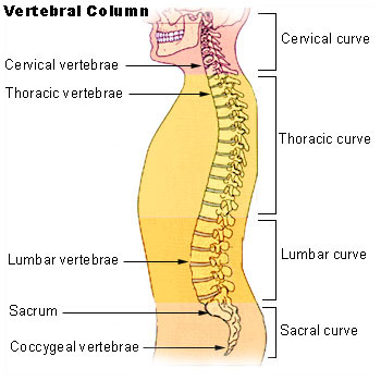 Vertebral column.
 -
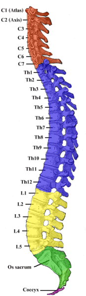 Vertebral column.
 -
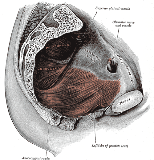 Left Levator ani from within.
 -
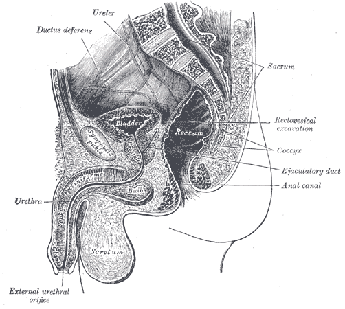 Median sagittal section of male pelvis.
 -
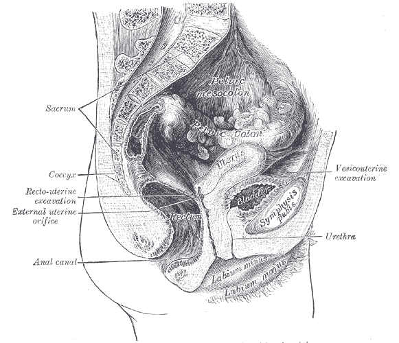 Median sagittal section of female pelvis.


See also
- Coccydynia (coccyx pain, tailbone pain)
- Bone terminology
- Terms for anatomical location
- Ganglion impar
References
- ↑ Coccyx, Dorland's Illustrated Medical Dictionary
- ↑ Idiopathic coccygodynia. Analysis of fifty-one operative cases and a radiographic study of the normal coccyx. The Journal of bone and joint surgery. American volume. 1983 Oct; 65(8): 1116-1124. Postacchini F, Massobrio M
- ↑ Clinical and radiological differences between traumatic and idiopathic coccygodynia. Yonsei Medical Journal, 1999 Jun, 40:3, 215-20. Kim NH; Suk KS
- ↑ Maigne JY, Molinie V, Fautrel B. (1992). "Anatomie des disques coccygiens". Revue de Médecine Orthopedique. 28: 34–35.
- ↑ Saluja PG. (1988). "The incidence of ossification of the sacrococcygeal joint". Journal of Anatomy. 156: 11–15.
- ↑ Causes and Mechanisms of Common Coccydynia. Spine, 2000, volume 25, number 23, 3072-3079. Maigne, J-Y, Doursounian, L, Chatellier, G.
- ↑ Foye P, Buttaci C, Stitik T, Yonclas P (2006). "Successful injection for coccyx pain". Am J Phys Med Rehabil. 85 (9): 783–4. PMID 16924191.
External links
- Template:GraySubject
- Template:SUNYAnatomyLabs - "The Female Perineum: Osteology"
- Template:ViennaCrossSection
- Coccyx pain: diagnosis, coping and treatment at coccyx.org
- coccyx-fracture(Tailbone Fracture) at al-hikmah.org
- Coccydynia (coccyx pain, tailbone pain) at eMedicine (Peer-reviewed medical chapter, available free online)
- For more information on Dr. Foye's treatments for Tailbone Pain please see: www.TailboneDoctor.com Note that medical advice can not be given to patients who have not yet been seen by Dr. Foye in his office.
bg:Опашна кост ca:Còccix de:Steißbein eo:Kokcigo it:Coccige lt:Uodegikaulis nl:Stuit no:Haleben sk:Kostrč sl:Trtica fi:Häntäluu uk:Куприкова кістка