Cardia
Editor-In-Chief: C. Michael Gibson, M.S., M.D. [1]
Overview
|
WikiDoc Resources for Cardia |
|
Articles |
|---|
|
Media |
|
Evidence Based Medicine |
|
Clinical Trials |
|
Ongoing Trials on Cardia at Clinical Trials.gov Clinical Trials on Cardia at Google
|
|
Guidelines / Policies / Govt |
|
US National Guidelines Clearinghouse on Cardia
|
|
Books |
|
News |
|
Commentary |
|
Definitions |
|
Patient Resources / Community |
|
Directions to Hospitals Treating Cardia Risk calculators and risk factors for Cardia
|
|
Healthcare Provider Resources |
|
Continuing Medical Education (CME) |
|
International |
|
|
|
Business |
|
Experimental / Informatics |
The cardia (or esophagogastric junction [1][2] or gastroesophageal junction [3][4][5][6]) is the anatomical term for the junction orifice of the stomach and the esophagus. At the cardia, the mucosa of the esophagus transitions into gastric mucosa.
The cardia is also the site of the lower esophageal sphincter (LES)[7][8][9] (also termed cardiac sphincter[10], gastroesophageal sphincter[11], and esophageal sphincter[12]).
Nomenclature and classification
There is disagreement in the academic anatomy community over whether the cardia is part of the stomach, part of the esophagus or a distinct entity, as described in this article. The difference is more than semantic when used in clinical studies and applied to individual patients.
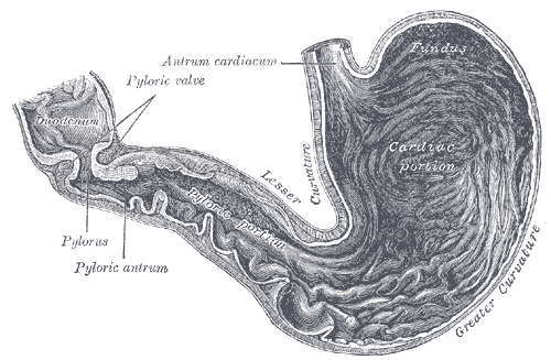
Classical anatomy textbooks, and some other resources[13][14], describe the cardia as the first of 4 regions of the stomach. This makes sense histologically because the mucosa of the cardia is the same as that of the stomach.
Many recent writings describe it as the LES.
Function

The stomach generates strong acids and enzymes to aid in food digestion. This digestive mixture is called gastric juice. The inner lining of the stomach has several mechanisms to resist the effect of gastric juice on itself, but the mucosa of the esophagus does not. The esophagus is normally protected from these acids by a one-way valve mechanism at its junction with the stomach. This one-way valve is called the lower esophageal sphincter (LES), and prevents gastric juice from flowing back into the esophagus.
During peristalsis, the LES allows the food bolus to pass into the stomach. It prevents chyme, a mixture of bolus, stomach acid, and digestive enzymes, from returning up the esophagus. The LES is aided in the task of keeping the flow of materials in one direction by the diaphragm.
Histology
On histological examination, the junction can be identified by the following transition:[15][16]
- nonkeratinized stratified squamous epithelium in the esophagus
- simple columnar epithelium in the stomach
However, in Barrett's esophagus, the epithelial distinction may vary, so the histological border may not be identical with the functional border.
The cardiac glands can be seen in this region. They can be distinguished from other stomach glands (fundic glands and pyloric glands) because the glands are shallow and simple tubular.
Pathology
Deficiencies in the strength or the efficiency of the LES lead to various medical problems involving acid damage on the esophagus.
In achalasia, one of the defects is failure of the LES to relax properly.
Etymology
The word comes from the Greek kardia meaning heart, the cardiac orifice of the stomach.
Additional images
-
-
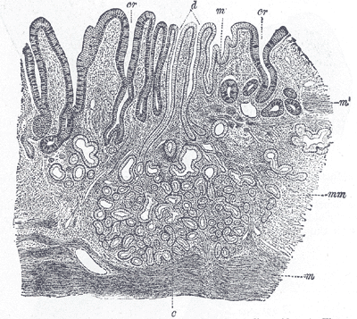
Section of mucous membrane of human stomach, near the cardiac orifice. X 45.


References
- ↑ Template:EMedicineDictionary
- ↑ Template:Dorlands
- ↑ Template:LoyolaMedEd
- ↑ http://cellbio.utmb.edu/microanatomy/digestive/Esophagus.htm
- ↑ http://www.cancer.org/docroot/cri/content/cri_2_4_1x_what_is_esophagus_cancer_12.asp?sitearea=cri
- ↑ http://www.cancer.gov/Templates/db_alpha.aspx?CdrID=302458
- ↑ http://hopkins-gi.nts.jhu.edu/pages/latin/templates/index.cfm?pg=disease1&organ=1&disease=13&lang_id=1
- ↑ http://www.physio.unr.edu/ICC/gallery/LES-1.htm
- ↑ med/2965 at eMedicine
- ↑ Template:EMedicineDictionary
- ↑ Template:Dorlands
- ↑ http://www.mcg.edu/Otolaryngology/patientGERD.htm
- ↑ Template:Dorlands
- ↑ Template:SUNYAnatomyLabs - "Abdominal Cavity: The Stomach"
- ↑ Histology image: 11101loa – Histology Learning System at Boston University
- ↑ Histology image: 11111ooa – Histology Learning System at Boston University