Brachial plexus
Template:Infobox Nerve Editor-In-Chief: C. Michael Gibson, M.S., M.D. [1]; Associate Editor(s)-in-Chief:
|
WikiDoc Resources for Brachial plexus |
|
Articles |
|---|
|
Most recent articles on Brachial plexus Most cited articles on Brachial plexus |
|
Media |
|
Powerpoint slides on Brachial plexus |
|
Evidence Based Medicine |
|
Clinical Trials |
|
Ongoing Trials on Brachial plexus at Clinical Trials.gov Trial results on Brachial plexus Clinical Trials on Brachial plexus at Google
|
|
Guidelines / Policies / Govt |
|
US National Guidelines Clearinghouse on Brachial plexus NICE Guidance on Brachial plexus
|
|
Books |
|
News |
|
Commentary |
|
Definitions |
|
Patient Resources / Community |
|
Patient resources on Brachial plexus Discussion groups on Brachial plexus Patient Handouts on Brachial plexus Directions to Hospitals Treating Brachial plexus Risk calculators and risk factors for Brachial plexus
|
|
Healthcare Provider Resources |
|
Causes & Risk Factors for Brachial plexus |
|
Continuing Medical Education (CME) |
|
International |
|
|
|
Business |
|
Experimental / Informatics |
Overview
The brachial plexus is an arrangement of nerve fibres, running from the spine, specifically from above the fifth cervical vertebra to underneath the first thoracic vertebra (C5-T1). It proceeds through the neck, the axilla (armpit region) and into the arm.
Function
- The brachial plexus is responsible for cutaneous and muscular innervation of the entire upper limb, with two exceptions: the trapezius muscle innervated by the spinal accessory nerve and an area of skin near the axilla innervated by the intercostobrachialis nerve.
- This function may be impaired by tumor growth of the Apical region of either Lung.
- Therefore, brachial plexus lesions can lead to severe functional impairment.
Anatomy
One can remember the order of brachial plexus elements by way of the mnemonic, "Read The Damn Cadaver Book" - Roots, Trunks, Divisions, Cords, Branches or - Roots, Trunks, Divisions, Cords, Collateral/Pre-terminal Branches, and (Terminal) Branches.
- The five roots are the five anterior rami of the spinal nerves, after they have given off their segmental supply to the muscles of the neck.
- These roots merge to form three Trunks:
- Superior or Upper (C5-C6)
- Middle (C7)
- Inferior or Lower (C8-T1)
- Each trunk then splits in two, to form six Divisions:
- Anterior division of the superior, middle and inferior trunks
- Posterior division of the superior, middle and inferior trunks
- These six divisions will re-group to become the three Cords. The cords are named by their position in respect to the axillary artery.
- The posterior cord is formed from the three posterior divisions of the trunks (C5-T1).
- The lateral cord is the anterior divisions from the upper and middle trunks (C5-C7).
- The medial cord is simply a continuation of the lower trunk (C8-T1).
- The branches are listed below. Most branch off of the cords, but a few branch (indicated in italics) directly off of earlier structures. The five in bold are considered "terminal branches".
Diagram

Specific branches
| From | Nerve | Roots | Muscles | Cutaneous | Diseases associated |
| roots | dorsal scapular nerve | C5 | rhomboid muscles and levator scapulae | - | |
| roots | long thoracic nerve | C5, C6, C7 | serratus anterior | - | |
| superior trunk | nerve to the subclavius | C5, C6 | subclavius muscle | - | |
| superior trunk | suprascapular nerve | C5, C6 | supraspinatus and infraspinatus | - | |
| lateral cord | lateral pectoral nerve | C5, C6, C7 | pectoralis major (by communicating with the medial pectoral nerve) | - | |
| lateral cord | musculocutaneous nerve | C5, C6, C7 | coracobrachialis, brachialis and biceps brachii | becomes the lateral cutaneous nerve of the forearm | |
| lateral cord | lateral root of the median nerve | C5, C6, C7 | fibres to the median nerve | - | |
| posterior cord | upper subscapular nerve | C5, C6 | subscapularis (upper part) | - | |
| posterior cord | thoracodorsal nerve | C6, C7, C8 | latissimus dorsi | - | |
| posterior cord | lower subscapular nerve | C5, C6 | subscapularis (lower part) and teres major | - | |
| posterior cord | axillary nerve | C5, C6 | anterior branch: deltoid and a small area of overlying skin posterior branch: teres minor and deltoid muscles |
posterior branch becomes upper lateral cutaneous nerve of the arm | |
| posterior cord | radial nerve | C5, C6, C7, C8, T1 | triceps brachii, supinator, anconeus, the extensor muscles of the forearm, and brachioradialis | skin of the posterior arm as the posterior cutaneous nerve of the arm | |
| medial cord | medial pectoral nerve | C8, T1 | pectoralis major and pectoralis minor | - | |
| medial cord | medial root of the median nerve | C8, T1 | fibres to the median nerve | portions of hand not served by ulnar or radial | |
| medial cord | medial cutaneous nerve of the arm | C8, T1 | - | front and medial skin of the arm | |
| medial cord | medial cutaneous nerve of the forearm | C8, T1 | - | medial skin of the forearm | |
| medial cord | ulnar nerve | C8, T1 | flexor carpi ulnaris, the medial 2 bellies of flexor digitorum profundus, most of the small muscles of the hand | the skin of the medial side of the hand and medial one and a half fingers on the palmar side and medial two and a half fingers on the dorsal side |
Additional images

-
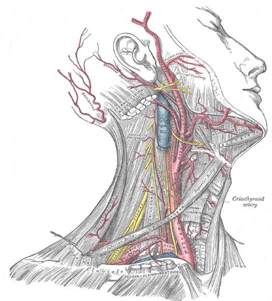
Superficial dissection of the right side of the neck, showing the carotid and subclavian arteries.
-

The axillary artery and its branches.
-
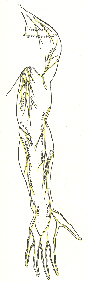
Cutaneous nerves of right upper extremity. Posterior view.
-
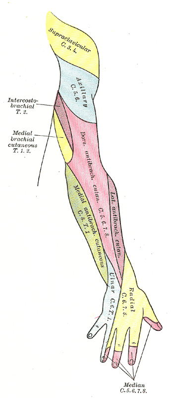
Diagram of segmental distribution of the cutaneous nerves of the right upper extremity. Posterior view.
-
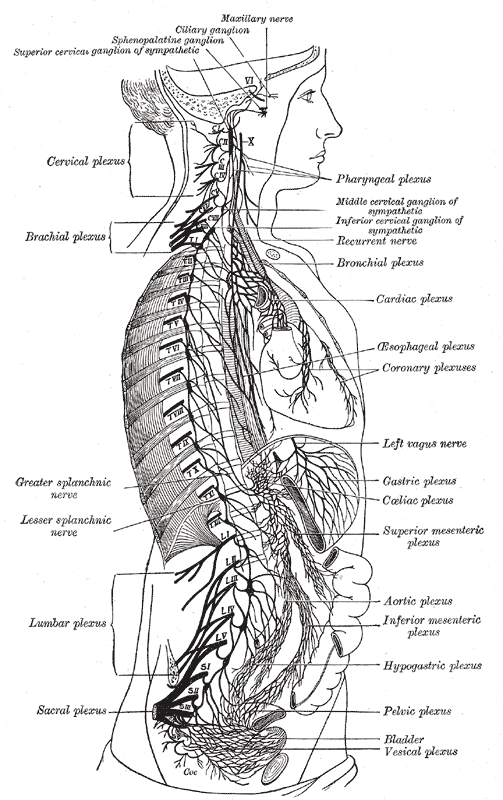
The right sympathetic chain and its connections with the thoracic, abdominal, and pelvic plexuses.
-

Side of neck, showing chief surface markings.


See also
References
External links
Template:Spinal nerves Template:Brachial plexus
de:Plexus brachialis hr:Ručni splet id:Plexus brachialis he:מקלעת הזרוע no:Plexus brachialis sv:Plexus brachialis