Arytenoid cartilage
Overview
The arytenoid cartilages are a pair of small three-sided pyramids which form part of the larynx, to which the vocal cords are attached.
Each is pyramidal in form, and has three surfaces, a base, and an apex.
Surfaces
The posterior surface is a triangular, smooth, concave, and gives attachment to the Arytænoidei obliquus and transversus.
The antero-lateral surface is somewhat convex and rough. On it, near the apex of the cartilage, is a rounded elevation (colliculus) from which a ridge (crista arcuata) curves at first backward and then downward and forward to the vocal process. The lower part of this crest intervenes between two depressions or foveæ, an upper, triangular, and a lower oblong in shape; the latter gives attachment to the Vocalis muscle.
The medial surface is narrow, smooth, and flattened, covered by mucous membrane, and forms the lateral boundary of the intercartilaginous part of the rima glottidis.
Base and apex
The base of each cartilage is broad, and on it is a concave smooth surface, for articulation with the cricoid cartilage.
- Its lateral angle is short, rounded, and prominent; it projects backward and lateralward, and is termed the muscular process; it gives insertion to the posterior cricoarytenoid muscles behind, and to the lateral cricoarytenoid muscles in front.
- Its anterior angle, also prominent, but more pointed, projects horizontally forward; it gives attachment to the vocal ligament, and is called the vocal process.
The apex of each cartilage is pointed, curved backward and medialward, and surmounted by a small conical, cartilaginous nodule, the corniculate cartilage.
Function
They allow the vocal cords to be tensed, relaxed, or approximated.
The arytenoids articulate with the supero-lateral parts of the cricoid cartilage lamina, forming the cricoarytenoid joints at which they can come together, move apart, tilt anteriorly or posteriorly, and rotate.
Additional images
-
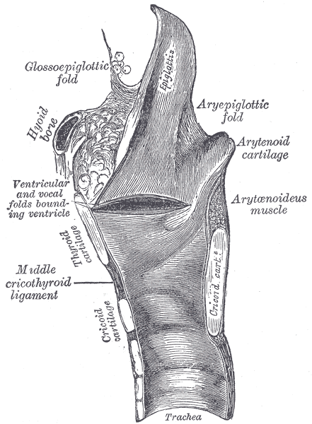
Sagittal section of the larynx and upper part of the trachea.
-
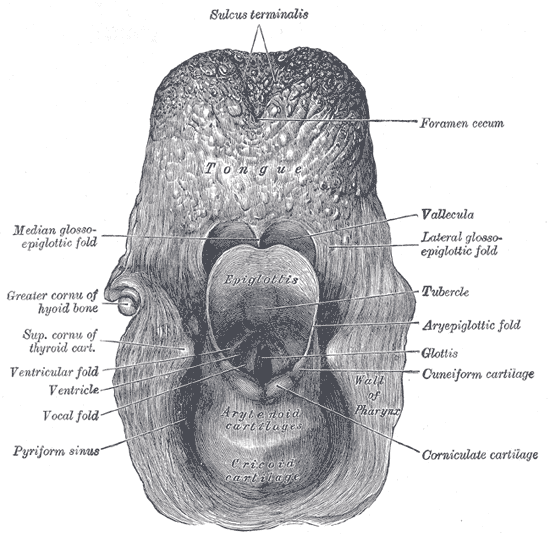
The entrance to the larynx, viewed from behind.
-
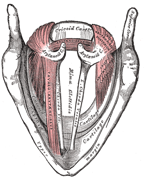
Muscles of the larynx, seen from above.


